Cell by Cell: How Dapagliflozin Rewires the Kidney
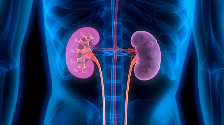
Key Takeaway: For the first time, researchers using serial kidney biopsies and single-cell RNA sequencing in patients on an SGLT2 inhibitor have revealed that dapagliflozin reprograms kidney cells at the molecular level. This reprogramming mitigates glycolysis, oxidative stress, inflammation, and fibrosis while reinforcing cellular structural integrity. These tissue-level changes parallel clinical improvements in renal filtration and oxygenation, offering the most detailed evidence to date on how this drug class protects the kidneys.
A Window into the Living Kidney
For years, we have known that SGLT2 inhibitors have a remarkable effect on the kidneys. Clinical trials involving tens of thousands of patients have demonstrated that these drugs reduce kidney failure, hospitalizations, and cardiovascular deaths[2]. But there is a vast difference between knowing that a drug works and understanding how it works. Until now, the molecular mechanisms behind the kidney-protective effects of SGLT2 inhibitors were pieced together from animal models and indirect biomarkers—data projected, in a sense, onto the black box of human tissue. For the first time, scientists have looked inside the kidneys of patients taking an SGLT2 inhibitor, and what they found at the single-cell level is a masterpiece of mechanistic elucidation.
Study Methodology
This was a placebo-controlled trial conducted in young people with type 1 diabetes who showed signs of hyperfiltration—a condition where the kidneys are working under excessive pressure, a well-known early step on the path to diabetic kidney disease[3]. Participants were given either dapagliflozin or a placebo. What made the study extraordinary was the use of serial kidney biopsies in adults aged 18 and over. This means tissue was sampled at multiple time points, allowing researchers to observe changes within the same organ over time.
The biopsy samples were then analyzed using single-cell RNA sequencing technology. This technology reads the genetic activity of individual cells rather than averaging the signals across an entire piece of tissue. The result was a dataset of 214,415 individual cells, each cataloged by its type and transcriptional profile[1]. Alongside the biopsies, the team performed renal MRI imaging to assess tissue oxygenation and collected urine for proteomic analysis, creating a multi-layered picture spanning from molecule to organ.
Findings: A Cellular-Level Investigation
The findings resemble a coordinated rescue operation being carried out simultaneously across multiple cell types in the kidney.
- Proximal tubule cells—the workhorses of the kidney responsible for reabsorbing glucose, electrolytes, and water—showed a marked down-regulation in the expression of markers for glycolysis and oxidative stress. In diabetic kidneys, these cells are chronically overloaded. Excess glucose floods the tubular system, forcing the cells into metabolic overdrive, which generates toxic reactive oxygen species[4]. By blocking SGLT2 transporters right in these cells, dapagliflozin reduced the metabolic burden at its source.
- Endothelial cells, which line the kidney’s intricate vascular network, exhibited reduced expression of pro-fibrotic and inflammatory genes. This is critically important, as microvascular damage and progressive scar tissue formation (fibrosis) are hallmarks of advanced diabetic nephropathy[5].
- Podocytes, the specialized cells that wrap around the glomerular capillaries and maintain the kidney’s filtration barrier, showed a reinforcement of their cytoskeleton. Podocyte loss is an essentially irreversible process and a strong predictor of progression to kidney failure. This finding that dapagliflozin supports the structural framework of these cells points to a protective effect at one of the kidney’s most vulnerable points.
These molecular changes were not isolated laboratory findings. They paralleled measurable clinical improvements: a reduction in hyperfiltration and normalization of medullary oxygenation on MRI. The kidney’s inner medulla operates under near-hypoxic conditions even in a healthy state; in diabetes, oxygen delivery worsens, accelerating tubular damage[6]. The restoration of oxygenation observed in this study suggests that the metabolic reprogramming by dapagliflozin translates into a tangible reduction in tissue stress.
Adding another layer of confirmation, urinary proteomic analysis mirrored the tissue-level changes. Damage markers decreased while protective proteins increased. This could one day become a non-invasive signature to help clinicians monitor drug response without the need for a biopsy needle.
The Mechanism: Why Metabolic Reprogramming Matters
To grasp the significance of these findings, one must consider the fundamental problem in the diabetic kidney. Chronically high glucose overwhelms the reabsorptive capacity of the proximal tubule. The cell responds by ramping up glycolytic enzymes and mitochondrial activity, which produces reactive oxygen species as a byproduct. These free radicals damage DNA, proteins, and lipid membranes, triggering inflammatory cascades that call in immune cells and activate fibroblasts[4]. Over years, this cascade converts functional nephrons into scar tissue.
SGLT2 inhibitors interrupt this process at its very beginning. By blocking the sodium-glucose cotransporter 2 in the S1 and S2 segments of the proximal tubule, dapagliflozin reduces the glucose load these cells have to handle. The downstream consequences—less glycolysis, fewer reactive oxygen species, calmer inflammatory signaling, and decreased fibrotic gene expression—are now supported by evidence at the single-cell level. The podocyte findings add a dimension that animal models had hinted at but had never been confirmed in human tissue: structural reinforcement of the filtration barrier itself.
Study Limitations
While this study is groundbreaking, its context is important. The study group consisted of young patients with type 1 diabetes, who differ metabolically and immunologically from the older adults with type 2 diabetes who make up the majority of SGLT2 inhibitor prescriptions. Whether the exact same transcriptional changes would occur in a 65-year-old with long-standing type 2 diabetes and established nephropathy has yet to be shown. The sample size, inherently limited by the ethical and logistical challenges of serial kidney biopsies, constrains the statistical power for subgroup analyses. While the convergence of biopsy, MRI, and proteomic data is compelling, a single study is not definitive proof; replication in larger, more diverse populations will be essential.
Clinical Implications
For the millions of patients with diabetes already taking an SGLT2 inhibitor, this study offers something rare: a mechanistic explanation for the protection they are likely already seeing. These findings validate the biological rationale for starting these drugs early, before fibrosis sets in and podocyte loss reaches a point of no return. For clinicians, the urinary proteomic signatures identified here could serve as real-time biomarkers of drug efficacy in the future, allowing for personalized dosing or combination strategies. For the field of nephrology at large, this work sets a new standard: if you want to understand how a drug protects the kidney, you have to look inside it—cell by cell. That is not always easy to do, but this study has achieved it.
Scientific Sources
- Bjornstad P, et al. SGLT2 inhibition modulates metabolic, vascular, and inflammatory molecular markers in the kidney in youth with type 1 diabetes. Science translational medicine. 2026;18(859):eaee1005. PubMed: https://pubmed.ncbi.nlm.nih.gov/42485434/
- Heerspink HJL, et al. Dapagliflozin in Patients with Chronic Kidney Disease. N Engl J Med. 2020. DOI: 10.1056/NEJMoa2024816
- Helal I, et al. Glomerular hyperfiltration: definitions, mechanisms and clinical implications. Nat Rev Nephrol. 2012. DOI: 10.1038/nrneph.2012.19
- Forbes JM, et al. Oxidative stress as a major culprit in kidney disease in diabetes. Diabetes. 2008. DOI: 10.2337/db08-0057
- Kanwar YS, et al. Diabetic nephropathy: mechanisms of renal disease progression. Exp Biol Med. 2008. DOI: 10.3181/0705-MR-134
- Friederich-Persson M, et al. Kidney hypoxia, attributable to increased oxygen consumption, induces nephropathy independently of hyperglycemia and oxidative stress. Kidney Int. 2013. DOI: 10.1161/HYPERTENSIONAHA.113.01425
Medically reviewed by
Dr. Şekip Altunkan
Dr. Şekip Altunkan is an internal medicine specialist with extensive clinical experience. He trained at Hacettepe University Faculty of Medicine and later served as an Associate Professor in Internal Medicine. He founded and led the Metropol Internal Medicine and Hypertension Clinic in Ankara, pioneering non-invasive Electron Beam Tomography (EBT) cardiac imaging, arterial-stiffness measurement, and nationwide Holter monitoring. He currently practices at his private clinic in Ankara, focusing on hypertension, vascular health, cholesterol, diabetes and heart disease. He has published widely in national and international journals, serves as a peer reviewer for several international journals, and is the author of the book "Questions and Answers on Hypertension."