Crystallized Agony: The Cellular Warfare Behind Gout and the Uric Acid Illusion
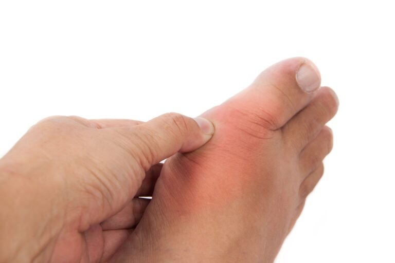
Key Takeaway: Modern metabolic medicine and immunology demonstrate that gout is a complex, systemic inflammatory disorder rather than a superficial consequence of dietary overindulgence, driven by a hominid evolutionary lack of the uricase enzyme and a physicochemical saturation threshold for monosodium urate (MSU) crystals (approximately 6.8 mg/dL). When serum levels overflow this threshold, needle-like MSU crystals precipitate in peripheral, cooler joints—specifically the first metatarsophalangeal joint—where they act as danger-associated molecular patterns, rupturing macrophage phagolysosomes and detonating the volatile NLRP3 inflammasome / IL-1 Beta cytokine storm. Crucially, clinicians must avoid the common diagnostic trap of treating hyperuricemia as synonymous with acute gout, and adhere to the strict golden rule: never initiate uric acid-lowering therapies like Allopurinol during an active flare, as the resulting crystal-shedding phenomenon violently intensifies the inflammatory war zone. Long-term victory requires managing the patient’s complete cardiometabolic and renal profile, slowly titrating medications to maintain serum urate permanently below 6 mg/dL, and shifting the nutritional focus away from obsolete purine-banning dogmas toward aggressively eliminating true metabolic super-triggers like industrial high-fructose corn syrup and guanosine-rich beer.
Introduction: From the Disease of Kings to a Cellular Energy Crisis
Medical history thoroughly enjoys branding certain pathologies with social classes and structures of power. The oldest, most dramatic, and most popular victim of this historical labeling in medical literature is undoubtedly Gout. For centuries, from Alexander the Great to Henry VIII, spanning a vast elite demographic of emperors of collapsing empires and wealthy bourgeois, this clinical presentation was long hailed as the “Disease of Kings” or the “King of Diseases”. The classical narrative superficially reduced this condition to a mere consequence of protein excess, explicitly linking it to overindulgence in red meat, a luxurious lifestyle, and unrestricted alcohol consumption. Modern metabolic medicine and immunology, however, whisper a far deeper, far more insidious molecular reality.
Gout is not merely a transient ache in the big toe following a lavish Sunday feast. It must be recognized as a systemic, chronic, and inflammatory metabolic crisis—one that originates in the deep pathways of purine metabolism, is fueled by genetic architecture, occupies intracellular signaling networks, and drives the immune system into an absolute civil war. Globally, the prevalence of gout ranges from 1% to 7%, presenting significantly higher rates in men than in women, though a notable increase is observed in postmenopausal women. Crucially, gout is never confined strictly to the joints; it must be evaluated as a systemic metabolic disorder intimately intertwined with a wide spectrum of comorbidities, including cardiovascular disease, chronic kidney disease (CKD), diabetes, metabolic syndrome, and hypertension.
In this comprehensive edition of The Vault, we will dismantle the deceptive illusion created by raw uric acid values, expose the microscopic crystal landmines accumulating silently within joint spaces, navigate the diagnostic traps that fiercely challenge clinicians in emergency departments and clinics, and map the modern therapeutic revolution extending from classical modalities to advanced biologic agents.
Molecular Pathophysiology: The Purine Matrix and Crystallization Dynamics
To comprehend the pathophysiological universe of gout, one is compelled to descend to the most intimate building blocks of the cell nucleus: the purine bases (adenine and guanine) that form the structural foundation of DNA and RNA. In the human body, purines are generated endogenously through the breakdown of aging cellular matter and introduced exogenously via dietary intake. The ultimate metabolic waste product of this continuous purine traffic is the uric acid molecule.
At this juncture, human evolution executed a bizarre biological paradox. The vast majority of mammals possess the enzyme uricase (urate oxidase), which seamlessly converts uric acid into allantoin—a highly water-soluble compound easily excreted by the body. However, during hominid evolution millions of years ago, a pivotal genetic mutation rendered humans entirely incapable of synthesizing this enzyme. Consequently, we are left with a biological blueprint naturally prone to crystallization; we must excrete raw, unaltered uric acid directly through our kidneys and gastrointestinal tract.
The Uric Acid Illusion: The Chasm Between Hyperuricemia and Gout
In clinical practice, the most common trap into which both patients and inexperienced physicians fall is conflating elevated uric acid (hyperuricemia) with clinical gout. A laboratory report revealing a serum uric acid level of 8 mg/dL does not automatically render that patient a gout patient. Hyperuricemia represents a mandatory pre-condition for the development of gout, yet it is not a standalone diagnosis. Only a small percentage of individuals with asymptomatic hyperuricemia will ever experience a clinical, acute gout flare in their lifetime.
When and why, then, does this dissolved uric acid transform into a vascular weapon? This transition is governed strictly by the physicochemical solubility threshold of Monosodium Urate (MSU) crystals. In human plasma, at a core body temperature of 37°C, the saturation threshold for uric acid is approximately 6.8 mg/dL. Once this boundary is breached, the solution becomes supersaturated, and uric acid begins to precipitate, combining with sodium ions to form needle-like, razor-sharp monosodium urate crystals.
Why Crystals Select the First Metatarsophalangeal (1st MTP) Joint
The classical onset of an acute gout attack in the big toe (podagra) is dictated by three elegant physical mechanisms rather than mere coincidence:
- Temperature Drop: The body’s peripheral extremities are inherently cooler than the core temperature. As temperature declines, the solubility of uric acid in synovial fluid drops dramatically, exponentially accelerating the rate of crystal precipitation.
- Microtrauma and Mechanical Stress: The constant weight-bearing and mechanical microtraumas experienced by the big toe during ambulation cause a steady shedding of proteoglycans from the joint cartilage. This structural shedding creates perfect focal points (nucleation sites) for crystals to adhere and accumulate.
- Intra-Articular Fluid Resorption: During nocturnal rest, water from the joint space is gradually reabsorbed back into the vascular system ; however, uric acid molecules remain physically trapped within the joint capsule. The secret behind that agonizing, sudden midnight gout flare is this abrupt, nocturnal spike in intra-articular uric acid concentration.
The Cellular Detonator: NLRP3 Inflammasome Activation
The needle-shaped MSU crystals that settle into the joint space are recognized by the innate immune system as dangerous foreign invaders, specifically classified as Danger-Associated Molecular Patterns (DAMPs). Resident macrophages patrolling the joint tissue attempt to engulf these crystals via phagocytosis. However, the crystals are so sharp and rigid that they physically rupture the macrophage’s internal digestive sacs (phagolysosomes), causing a violent intracellular explosion.
This cellular rupture awakens one of the most volatile molecular complexes in immunology: the NLRP3 Inflammasome. Once activated, the inflammasome triggers the caspase-1 enzyme, which cleaves sleeping intracellular precursors to release Interleukin-1 Beta (IL-1 Beta)—the most potent pro-inflammatory master cytokine in nature. IL-1 Beta aggressively dilates surrounding vessel walls, issues a systemic emergency alarm, and recruits thousands of circulating neutrophils into the joint space. Upon entry, these neutrophils launch a suicidal counter-offensive known as NETosis (Neutrophil Extracellular Traps). The result is absolute molecular chaos within the joint: the tissue burns, swells violently, and creates that legendary gout attack where the patient cannot tolerate even the weight of a bedsheet.
The Diagnostic Labyrinth: Avoiding Million-Dollar Mistakes
When an acute, erythematous, edematous, and entirely non-weight-bearing monoarthritis presents in a clinical examination room, the differential diagnosis list serves as a critical test of a physician’s acumen. Gout is a master masquerader; a misdiagnosis can lead to irreversible joint destruction.
Septic Arthritis: The Perilous Danger That Must Never Be Missed
The clinical presentation of an acute gout flare—including systemic fever, intense erythema, peripheral leukocytosis, and elevated C-reactive protein (CRP)—is virtually identical to septic arthritis, a bacterial infection within the joint space. The gravest sin a clinician can commit is administering a corticosteroid injection into a septic joint under the assumption of “definite gout,” which aggressively fuels the underlying bacterial infection, or conversely, unnecessarily subjecting a gout patient to surgical joint washing in an operating room.
- The Gold Standard: The definitive differentiation relies on joint fluid aspiration (arthrocentesis) followed immediately by polarized light microscopy. Finding needle-shaped, brightly fluorescent, negatively birefringent crystals confirms gout. However, the aspirate must always be sent for Gram staining and culture; a clinician must never forget that the presence of gout crystals does not physically preclude a co-existing septic arthritis within the same joint.
Pseudogout (CPPD): A Case of Crystal Identity Confusion
Gout is frequently mimicked by Calcium Pyrophosphate Deposition disease (CPPD), colloquially termed pseudogout. Here, the culprit is not monosodium urate, but rather calcium pyrophosphate molecules settling into tissue.
- Key Distinctions: Pseudogout preferentially targets larger joints like the knees and wrists rather than the big toe. Under a polarized microscope, these crystals appear as rhomboid, weakly positively birefringent structures rather than needles. Furthermore, conventional radiographs reveal a distinct, thin white line of calcification over the joint cartilage, a classic sign known as chondrocalcinosis.
Cellulitis and Soft Tissue Infections
Gout flares that spread over the dorsum of the foot and ankle are routinely misdiagnosed as cellulitis. Patients are frequently subjected to days of unnecessary intravenous antibiotic therapy without any regression of the inflammation. A meticulous physical exam solves this trap by demonstrating that the exquisite tenderness and erythema are strictly demarcated along the anatomical joint line, rather than spreading diffusely through the subcutaneous tissue.
Clinical Perspective: “The Saturated Glass and the Cellular Landmine Field”
In my outpatient clinic, when I consult with despondent patients who have strictly avoided red meat, tomatoes, and seafood for years yet still suffer four to five severe gout attacks annually, I always use this clinical metaphor to reframe their understanding of the disease:
“Imagine a glass sitting before you, filled to the absolute brim with water. We steadily add teaspoons of sugar (representing uric acid) and stir. Initially, the sugar dissolves completely, turning invisible; it flows through the blood harmlessly, much like free uric acid. However, there comes a tipping point where adding even a single grain of sugar overloads the solution. The glass becomes fully saturated, cannot bear the burden, and that final grain causes a massive mass of crystals to precipitate and crash to the bottom of the glass.
This is precisely what has occurred within your joints; they have transformed into a cellular landmine field packed with sharp, needle-tipped microscopic crystals. Even if you completely eliminate meat from your diet, your body continues to manufacture its own internal purines by breaking down its own aging cells. While those sharp landmines lie dormant in the joint space, a minor physical stub of the toe, a drop in local temperature, or sudden dehydration causes your immune system’s soldiers (macrophages) to step squarely onto a mine. The mine detonates, cells scream out inflammatory alarms from within, and your joint instantly becomes an active war zone.
Our objective as physicians is not merely to extinguish the fire during an acute flare by masking the pain of the exploding mines. Our true victory lies in using the correct long-term medications to steadily drain the water from that glass, permanently driving serum uric acid levels below 6 mg/dL. This molecular decompression slowly dissolves and completely erases those crystal landmines from the joint tissue. Treating gout solely with pain relievers without clearing the underlying crystal field is akin to living on top of a volatile bomb, waiting for the next detonation.”
Therapeutic Strategies: From Extinguishing Acute Inflammation to Chronic Restoration
Gout management features some of the most definitive algorithms in medicine, yet it remains one of the most frequently mismanaged areas in clinical practice. The therapeutic strategy must be bifurcated into two strict, non-communicating compartments: Acute Flare Management and Chronic Urate-Lowering Therapy (ULT).
Acute Flare Management: Extinguishing the Fire
During an acute attack, the absolute mandate is to rapidly suppress the localized NLRP3 inflammasome and IL-1 Beta storm. Our clinical arsenal holds three classical and two modern weapons:
- Colchicine: A brilliant, ancient molecule that blocks intracellular microtubule polymerization, effectively arresting the migration of neutrophils into the joint war zone. Clinical Pearl: The obsolete, toxic practice of administering colchicine hourly until the patient develops severe diarrhea has been completely abandoned by modern medicine. The current gold standard dictates a rapid loading dose of 1.2 mg (two tablets) within the first 12 hours of flare onset, followed by 0.6 mg (one tablet) one hour later, maintaining a prophylactic dose of 0.6 mg once or twice daily. When initiated early, its efficacy is unparalleled.
- Non-Steroidal Anti-Inflammatory Drugs (NSAIDs): High-dose indomethacin, naproxen, or etoricoxib can rapidly douse the inflammatory flame. However, a strict review of the patient’s baseline renal function (creatinine clearance) and gastric ulcer history is mandatory before initiation.
- Corticosteroids: In patients suffering from chronic kidney disease—a very frequent comorbidity in the gout population—where both NSAIDs and colchicine are contraindicated, short-term systemic oral methylprednisolone or direct intra-articular corticosteroid injection is highly effective and kidney-safe.
The Most Critical Golden Rule for Clinicians: While an acute gout flare is actively ongoing, NEVER initiate a medication like Allopurinol to lower blood uric acid . Abruptly dropping serum uric acid levels during an active flare destabilizes the surface architecture of large intra-articular crystal deposits. This destabilization causes them to fracture and shed mass directly into the joint fluid (the shedding phenomenon), violently intensifying and indefinitely prolonging the attack. If a patient is already taking allopurinol when an attack occurs, the drug must be maintained at the exact same dose ; however, if it is a new initiation, one must wait at least 2 to 3 weeks until the flare has completely resolved.
Chronic Management (ULT): The Era of Crystal Dissolution
Once the acute inflammation has entirely cleared, the focus shifts to dissolving the landmines at the bottom of the glass. The clinical goal is to maintain serum uric acid permanently under 6 mg/dL (and under 5 mg/dL if the patient presents with palpable tophi—large, visible physical nests of aggregated crystals).
- Xanthine Oxidase Inhibitors (Allopurinol & Febuxostat): These agents shut down the metabolic factory that converts purines into uric acid by blocking the xanthine oxidase enzyme. Allopurinol is our frontline agent; however, it must be initiated at a conservative dose (100 mg/day) and titrated upward slowly based on renal function. Risk: In patients of specific ancestral descents carrying the HLA-B 5801 allele, allopurinol can trigger a fatal systemic cutaneous drug reaction known as DRESS syndrome. Febuxostat represents a highly potent alternative for patients who display allopurinol intolerance.
- Uricosuric Agents (Probenecid & Lesinurad): These medications block the URAT1 transport proteins in the renal proximal tubules, heavily increasing the excretion of uric acid through urine. They are strictly contraindicated in patients with a history of nephrolithiasis (kidney stones).
- The Biologic Revolution and Advanced Modalities:
- Pegloticase: A recombinant uricase enzyme reserved for severe, refractory cases where the entire body is occupied by extensive tophi that fail to dissolve with oral agents. It restores the evolutionary missing link externally, converting highly insoluble uric acid into highly soluble allantoin, completely melting decades of accumulated crystal burdens within months.
- Anakinra & Canakinumab: Biologic monoclonal antibodies targeting the IL-1 Beta molecule directly. In severe, refractory attacks where renal failure precludes the use of NSAIDs or steroids, these targeted therapies suppress the core engine of the inflammatory storm within hours.
Clinical Case Analysis: A Mismanagement Blueprint at a Medical Center
A 54-year-old male presented to my private clinic exhibiting profound edema, localized heat, erythema, and excruciating pain in his left first MTP joint. The patient was in such overwhelming agony that despite it being winter, he could not bear the physical friction of a woolen sock; he arrived at the clinic wearing a loose slipper on his left foot.
Deepening his medical history, we discovered that five days prior, he had visited a local medical center complaining of mild, early discomfort in his big toe. A laboratory panel run at that facility revealed a serum uric acid level of 9.2 mg/dL. The attending physician, stating, “Your uric acid is dangerously high, we must force it down immediately,” promptly prescribed Allopurinol 300 mg on the very first day of the acute flare. Within 24 hours of ingesting the first dose, the patient woke up to an unprecedented, unremitting wave of agony as the pain radiated across his entire foot. Furthermore, the patient’s medical background was highly complicated by uncontrolled hypertension and Stage 3 chronic kidney disease (Creatinine clearance: 38 mL/min).
Anatomy of Clinical Errors and the Resolution:
This case served as a perfect exhibition of the classic pitfalls in modern gout management:
- Error 1: Initiating a urate-lowering therapy (Allopurinol) in the absolute nadir of an acute flare destabilized the intra-articular crystal matrix, unleashing a massive shedding phenomenon that fueled the inflammatory storm.
- Error 2: In a patient with known Stage 3 chronic kidney disease, allopurinol was launched blindly at an aggressive dose of 300 mg without any baseline titration. In renal impairment, allopurinol must be initiated at exceptionally low doses (50 to 100 mg/day) and stepped up incrementally to mitigate the profound risk of severe systemic hypersensitivity and toxicity.
The Intervention:
I immediately suspended the allopurinol therapy. Given his severe renal impairment, loading him with high-dose NSAIDs or colchicine was entirely out of the question. Instead, I initiated a short, tightly controlled course of systemic corticosteroids to bypass the kidneys, combined with localized cold therapy.
The acute flare resolved completely within 48 hours. Once the patient was entirely pain-free and his first MTP joint returned to its natural anatomy, we waited exactly 3 weeks. We then cautiously reintroduced allopurinol—not at a blind 300 mg dose, but at a kidney-safe 50 mg/day, pairing it with low-dose colchicine (0.5 mg/day) prophylaxis to shield the joint from micro-crystal shedding during the shifting of the urate pool. Over the following months, we titrated the allopurinol dose step-by-step, comfortably steering his uric acid down to a stable 5.5 mg/dL without stressing his renal architecture. He has remained entirely free of a single gout attack for over a year.
As an internist who has spent decades managing metabolic diseases like hypertension, atherosclerosis, heart failure, and diabetes, I routinely encounter hyperuricemia and gout in my patient cohort. I must emphasize a critical clinical caveat here: certain diuretics used extensively to manage hypertension and heart failure can heavily reduce renal urate clearance, consequently elevating blood uric acid and precipitating violent gout flares. For a hypertensive patient with a known history of gout or a rising uric acid trajectory, clinicians should aggressively avoid these specific diuretics wherever possible.
However, clinical reality dictates that many advanced hypertensive profiles cannot be successfully controlled without a diuretic component, and diuretic therapy is a non-negotiable cornerstone in heart failure management. In these delicate, intertwined clinical scenarios, I want to clearly emphasize that rather than withholding vital cardiac protection, the physician must co-prescribe allopurinol alongside the diuretic to safely neutralize the metabolic backlash.
Furthermore, my long-term patients become master managers of their own gout profiles. Despite perfect medical compliance, occasional attacks can erupt due to unexpected systemic stressors. Because they are highly educated on their specific clinical parameters, they instantly contact me at the very first whisper of an attack. Recently, one of my patients caught me via phone at the airport just as he was boarding an international flight for an essential corporate summit abroad. The meeting was non-negotiable; he could not abort the trip. Because he was well-prepared, he had his rescue medications packed in his carry-on. Under my immediate remote guidance, we initiated his acute protocol right in his seat. He successfully completed his travel and delivered his summit entirely symptom-free.
The Diet Illusion: The Purine Trap, the Fructose Threat, and the Beer Reality
The most frequent question patients ask revolves around nutrition. The dietary management of uric acid and gout involves complex metabolic crossroads. The obsolete medical dogma of enforcing a draconian, unsustainable lifestyle by entirely banning red meat, tomatoes, and seafood has been completely discarded by contemporary metabolic medicine. Today, robust epidemiological data reveals that the rigid restriction of dietary purines yields a marginal reduction in serum uric acid of merely 1 to 2 mg/dL ; tracking a “matchbox-sized portion of meat” is mathematically incapable of preventing a saturated glass from overflowing.
The true, insidious enemy that must be aggressively combated is high-fructose corn syrup, heavily prevalent in processed foods and carbonated beverages. Fructose undergoes rapid phosphorylation in the liver, depleting cellular energy (ATP) and aggressively driving the intracellular synthesis of purines, turning on the uric acid production factory at full speed. On the alcohol front, the mechanism extends far beyond ethanol merely blocking the renal excretion of uric acid; beer specifically represents a concentrated, liquefied gout trigger due to its massive content of highly absorbable guanosine purines. While fermented beverages like wine exhibit a relatively neutral metabolic footprint when consumed in strict moderation, beer and high-proof spirits represent the most high-risk choices for igniting an intra-articular inflammatory storm. Thus, the modern clinician aims not to induce patient despair through starvation, but to eliminate “super-triggers” like beer and industrial fructose, relying on target-directed medical therapy to safely empty the saturated glass from the bottom up.
Conclusion: Crystal Management Beyond the Numbers
Gout is not a basic, mechanical biochemistry puzzle that can be solved simply by dragging laboratory numbers down on a screen. It represents a complex systemic intersection of intracellular inflammasome mechanics, evolutionary enzyme deficiencies, and vascular metabolic risks. Successful gout management demands that the clinician treat the patient’s kidneys, cardiovascular matrix, and metabolic risk factors (hypertension, obesity, diabetes) as a cohesive whole. Remember, the path to melting those hidden crystal landmines within the joints does not rely on starving the patient, but on flawlessly reading the molecular mechanisms driving the human organism.
Our Slogan: “Gout is not a mere disease of numbers restricted to a high uric acid reading; it is a cellular war fought over microscopic landmines buried deep within the joint. Quench the immediate flame calmly, but never declare victory until the landmines are entirely dissolved.”
Key Clinical Studies & Guidelines Reviewed
- Richette P, Bardin T. Gout. The Lancet. 2010;375(9711):318-328.
- FitzGerald JD, Dalbeth N, Mikuls TR, et al. 2020 American College of Rheumatology (ACR) Guideline for the Management of Gout. Arthritis & Rheumatology. 2020;72(6):879-895.
- Dalbeth N, Merriman TR, Stamp LK. Gout. The Lancet. 2016;388(10055):2039-2052.
- Timsans J, Palomäki A, & Kauppi M. Gout and Hyperuricemia: A Narrative Review of Their Comorbidities and Clinical Implications. Journal of Clinical Medicine. 2024;13(24):7616.
- Neogi T. Clinical Practice: Gout. New England Journal of Medicine. 2011;364(5):443-452.
- Major TJ, Topless RK, Dalbeth N, Merriman TR. Evaluation of the relative contribution of genetic profile and dietary factors to serum urate concentrations: a meta-analysis of population-based studies. BMJ. 2018;363:k3958.
- Emmerson BT. The Management of Gout. New England Journal of Medicine. 1996;334(7):445-451.
Medically reviewed by
Dr. Şekip Altunkan
Dr. Şekip Altunkan is an internal medicine specialist with extensive clinical experience. He trained at Hacettepe University Faculty of Medicine and later served as an Associate Professor in Internal Medicine. He founded and led the Metropol Internal Medicine and Hypertension Clinic in Ankara, pioneering non-invasive Electron Beam Tomography (EBT) cardiac imaging, arterial-stiffness measurement, and nationwide Holter monitoring. He currently practices at his private clinic in Ankara, focusing on hypertension, vascular health, cholesterol, diabetes and heart disease. He has published widely in national and international journals, serves as a peer reviewer for several international journals, and is the author of the book "Questions and Answers on Hypertension."