The Aneurysm Paradox: Fewer Ruptures, More Ticking
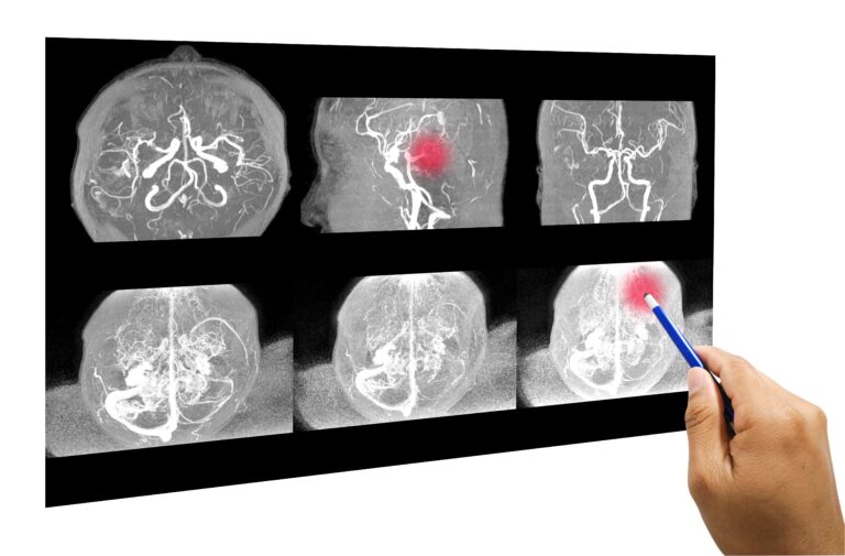
Key Takeaway: A large-scale global meta-analysis covering over 316,000 participants estimates that nearly 4 out of every 100 people carry an unruptured brain aneurysm, and this number is rising. Even as hemorrhagic stroke rates fall, the prevalence of larger, more dangerous aneurysms has doubled in the last decade, presenting a paradox that demands the urgent attention of clinicians and public health systems.
A Paradox in Plain Sight
We are facing a medical enigma that should unsettle anyone invested in stroke prevention. Over the past two decades, rates of subarachnoid hemorrhage—the catastrophic bleeding event that occurs when a brain aneurysm ruptures—have been declining in many parts of the world. This seems like progress. It feels like progress. But a massive new global analysis has revealed a starkly counterintuitive reality: the prevalence of aneurysms themselves is not falling, but rising. Moreover, the aneurysms becoming most common are the larger ones with the highest risk of one day rupturing.
What Did the Researchers Do?
This was no small undertaking. Investigators conducted a comprehensive meta-analysis encompassing 162 published papers and involving 316,131 participants from studies worldwide. The goal was as ambitious as it was simple: to produce the most reliable and up-to-date estimate of how common unruptured intracranial aneurysms (UIAs) truly are, identify who is most at risk, and track whether the prevalence has changed over time. The researchers pooled data from general population studies, high-risk cohorts, and autopsy series, applying rigorous statistical methods to account for differences in imaging technology, study design, and patient demographics.
What Did They Find?
The headline figure is this: in a reference population, the estimated prevalence of unruptured brain aneurysms was 3.9%[1]. This means roughly one in every 25 adults is walking around with a balloon-like weak spot on an artery inside their skull—most of them completely unaware.
But the data became far more alarming when the researchers looked at specific groups. In individuals with autosomal dominant polycystic kidney disease (ADPKD), the prevalence was 12.8%, while in those with connective tissue disorders like Ehlers-Danlos syndrome or Marfan syndrome, the rate was 10.3%. Among modifiable risk factors, being female carried a relative risk of 1.9, while hypertension was associated with a relative risk of 1.6 and current smoking with 1.4.
The layers of risk become even more distinct when focusing on the clinical anatomy and family history of this picture. More than half of the aneurysms (52.5%) were clustered in the carotid artery and its branches, with the middle cerebral artery (19.6%) and anterior cerebral artery (12.9%) being other common locations. In terms of size, nearly three-quarters (75.8%) of the detected lesions were small outpouchings under 5 millimeters. But the weight of genetic predisposition creates a critical threshold: the frequency of aneurysms was 6.7% in those with a single first-degree relative with an aneurysm or subarachnoid hemorrhage, but this rate jumped to 9.5% with two or more affected relatives. Furthermore, age creates independent momentum; while the prevalence is 2.9% in individuals under 55, it nearly doubles to 5.2% in those over 55, with each additional year of age corresponding to a relative increase of about 1.06% in aneurysm risk.
Perhaps the most striking finding was temporal. The prevalence of UIAs 5 millimeters or larger—the size threshold at which many neurosurgeons begin to seriously consider intervention—doubled from 0.7% in studies conducted between 2002 and 2015 to 1.4% in studies from 2016 to 2022. This is not a subtle shift. It’s a doubling in just over a decade.
The Mechanism: Why Arteries Betray Us
To understand why these numbers matter, you have to understand what a brain aneurysm actually is. Cerebral arteries have a three-layered structure: an inner lining (the intima), a muscular middle layer (the media), and an outer sheath (the adventitia). An aneurysm forms when the media degenerates at a point of hemodynamic stress—typically at a bifurcation where blood flow strikes the vessel wall at high velocity[2]. Over time, the wall thins, balloons outward, and creates a sac that fills with turbulent blood.
Hypertension accelerates this process by increasing the shear forces against an already vulnerable arterial wall. Smoking compounds the damage through chronic inflammation and the degradation of elastin and collagen, the structural proteins that keep vessel walls strong[3]. In patients with ADPKD, mutations in the PKD1 or PKD2 genes produce defective polycystin proteins, which are expressed not only in kidney tubules but also in vascular smooth muscle cells, weakening the arterial architecture from within[4]. Connective tissue disorders similarly undermine the extracellular matrix that gives arteries their resilience.
The nearly twofold higher incidence in women likely reflects the impact of postmenopausal estrogen withdrawal. Estrogen has vasculoprotective effects, promoting nitric oxide production and preserving the integrity of the arterial media[5]. When estrogen levels fall, the artery wall loses a critical line of defense against hemodynamic stress.
What’s Driving This Paradox?
So why are aneurysms becoming more common while subarachnoid hemorrhage rates are declining? Two explanations are most plausible. First, advanced and more widely available neuroimaging—especially high-resolution MR angiography—is detecting aneurysms that would have gone unnoticed a generation ago. Second, and more concerning, is that better acute stroke care and surgical management are preventing deaths from rupture without reducing the underlying formation of aneurysms. We may be getting better at managing the crisis, while the disease itself continues to spread.
A third, often overlooked possibility is the global demographic shift and environmental stressors. An analysis of nearly a century-long trend from 1931 to 2023 shows that aneurysm prevalence increased by an average of 2.5% per calendar year. However, this long-term increase loses its statistical power when adjusted for age, proving that the aging of the global population (and the growth of the over-65 demographic) is a primary driver in the accumulation of vascular weaknesses. Yet, the sharp increase observed within the 21st century (from 2.9% to 6.6% in healthy individuals screened with modern MRI/CT angiography between 2002–2015 and 2016–2022) cannot be explained solely by aging or the detection of small aneurysms with more sensitive devices. The absolute rate of aneurysms 5 millimeters and larger, which have a significant risk of bleeding, also doubled during this period (from 0.7% to 1.4%). This suggests that even as global rates of smoking and classic hypertension decline, new, not-yet-fully-understood metabolic, environmental, or vascular stressors continue to damage the vessel wall.
Notable Limitations
This meta-analysis inherits the limitations of its component studies: variable imaging protocols, different population demographics, and potential selection bias in cohorts undergoing brain imaging for other clinical reasons. The apparent rise in prevalence may partly reflect detection bias as imaging technology has improved. Additionally, many of the included studies come from East Asian populations, where aneurysm prevalence may differ from other ethnic groups for genetic reasons[6].
Another critical methodological dimension lies in the GRADE assessment, which rates the strength of the evidence. Since all included studies were observational, the overall level of evidence was rated as “low.” However, evidence from cohorts of healthy individuals, those with atherosclerosis, and those with polycystic kidney disease had “moderate” certainty, while data on connective tissue disorders and family history remained at a “very low” level of certainty. Furthermore, the data on connective tissue diseases had to group clinical entities like Ehlers-Danlos, Marfan, and Loeys-Dietz syndromes under a single umbrella, whereas the difference in aneurysm risk between cases with confirmed genetic mutations and individuals identified only phenotypically can vary significantly in clinical practice.
Conclusion: What These Findings Mean for Tomorrow
For patients with ADPKD or a connective tissue disorder, these data reinforce what many guidelines already suggest: routine screening with MR angiography should be part of the standard of care. For the broader population, the message is more nuanced but no less urgent. Hypertension and smoking remain the two most important modifiable risk factors. Every millimeter of mercury lowered, every cigarette not smoked, is a small act of structural preservation for the arteries that feed your brain.
For clinicians, the doubling in the prevalence of larger aneurysms requires us to recalibrate how aggressively we screen and how we counsel patients about incidental findings. The old assumption that falling rates of subarachnoid hemorrhage mean the aneurysm problem is shrinking is now obsolete. The aneurysms are out there. More of them are out there than before. And the conversation about what to do with that knowledge has never been more important.
This situation also presents a profound “overtreatment” dilemma for physicians and patients. With the frequency of aneurysms in healthy adults reaching roughly 1 in 15 (6.6%), and the majority of detected lesions being under 5 millimeters, interventions like surgical clipping or endovascular coiling carry their own morbidity risks. The fact that the number of detected aneurysms is skyrocketing while the global incidence of hemorrhage is decreasing indicates that the vast majority of these diagnosed sacs will remain silent and unruptured throughout a patient’s lifetime. Therefore, the new goal of modern medical practice is not to aggressively operate on every detected lesion. Instead, it is to develop a precise clinical surveillance strategy that avoids unnecessary procedural harm through tight blood pressure control, absolute smoking cessation, personalized PHASES risk scoring, and intermittent non-invasive imaging protocols.
Scientific Sources
- Dremel J, et al. Prevalence of unruptured intracranial aneurysms according to comorbidities, risk factors, country, and time period: a systematic review and meta-analysis. The Lancet. Neurology. 2026. PubMed: https://pubmed.ncbi.nlm.nih.gov/42679840/
- Stehbens WE. Etiology of intracranial berry aneurysms. J Neurosurg. 1989. DOI: 10.3171/jns.1989.70.6.0823
- Chalouhi N, et al. Review of cerebral aneurysm formation, growth, and rupture. Stroke. 2013. DOI: 10.1161/STROKEAHA.113.002390
- Pirson Y, et al. Intracranial aneurysms in autosomal dominant polycystic kidney disease. Kidney Int. 2002. DOI: 10.1038/ki.1994.151
- Mhurchu CN, et al. Hormonal factors and risk of aneurysmal subarachnoid hemorrhage. Stroke. 2001. DOI: 10.1161/01.str.32.3.606
- Vlak MH, et al. Prevalence of unruptured intracranial aneurysms, with emphasis on sex, age, comorbidity, country, and time period: a systematic review and meta-analysis. Lancet Neurol. 2011. DOI: 10.1016/S1474-4422(11)70109-0
Medically reviewed by
Dr. Şekip Altunkan
Dr. Şekip Altunkan is an internal medicine specialist with extensive clinical experience. He trained at Hacettepe University Faculty of Medicine and later served as an Associate Professor in Internal Medicine. He founded and led the Metropol Internal Medicine and Hypertension Clinic in Ankara, pioneering non-invasive Electron Beam Tomography (EBT) cardiac imaging, arterial-stiffness measurement, and nationwide Holter monitoring. He currently practices at his private clinic in Ankara, focusing on hypertension, vascular health, cholesterol, diabetes and heart disease. He has published widely in national and international journals, serves as a peer reviewer for several international journals, and is the author of the book "Questions and Answers on Hypertension."