The Brain Rusts for 16 Years Before Any Scan Sees It
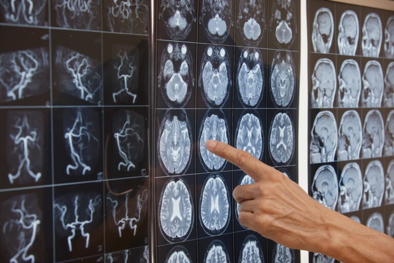
Key Takeaway: Advanced diffusion MRI technology can detect microscopic damage in the brain’s smallest blood vessels up to 16 years before standard imaging methods reveal any abnormalities. This extended silent period presents a groundbreaking opportunity for early intervention, potentially revolutionizing our paradigms for preventing stroke and dementia.
Invisible Erosion
Imagine a bridge slowly rusting from the inside out. For years, its surface paint looks flawless. Inspectors give it a clean bill of health. Then one day, with seemingly no warning, a girder buckles. The damage, of course, didn’t happen overnight; it accumulated silently, invisibly, for more than a decade. A strikingly similar process, it turns out, unfolds in the human brain—and a major new study reveals we finally have the tools to catch it.
The silent damage of cerebral small vessel disease—the progressive destruction of the brain’s smallest arteries and arterioles—can begin more than a decade before it becomes detectable on a conventional MRI scan. For millions of older adults told their brain imaging is ‘normal for their age,’ this may be a consolation delivered far too late.
How the Study Was Conducted
A team from the Mayo Clinic Study of Aging, one of the world’s most meticulously designed longitudinal brain health studies, followed 2,077 participants over time using two different types of brain imaging. The first was standard FLAIR-MRI, the bedrock of clinical practice that physicians rely on to detect white matter hyperintensities—bright spots indicating areas of tissue damage from chronic vascular disease. The second was advanced diffusion MRI, a technique that measures how water molecules move at a microscopic level within brain tissue. When the brain’s tissue architecture is intact, water molecules follow a predictable, orderly pattern. But when small blood vessels begin to fail and surrounding tissue starts to break down, these patterns shift in subtle but detectable ways.
By comparing these two imaging methods across repeated visits, the researchers could pinpoint when each scan type first registered abnormal findings and measure the time lag between them.
Key Findings
The results were significant. Diffusion MRI biomarkers of microstructural damage became abnormal 7 to 16 years before white matter hyperintensities became visible on standard scans[1]. A specific marker called the ‘Arteriolosclerosis-score,’ which specifically tracks damage to the walls of the brain’s tiniest arteries, reached abnormal values a full 16 years before standard MRI showed any changes in half the study population.
This implies the existence of a prolonged ‘pre-WMH’ (pre-white matter hyperintensity) phase of cerebral small vessel disease: a window of more than a decade when the brain is silently deteriorating, but conventional imaging sees nothing wrong. The study also found that cardiometabolic conditions like hypertension, diabetes, obesity, and high cholesterol predicted an earlier worsening across all biomarkers. Interestingly, the researchers observed sex-based differences in the timing of specific abnormalities—a finding that could inform more personalized screening strategies in the future.
The Mechanism: Why Are the Brain’s Smallest Vessels So Vulnerable?
To appreciate the significance, one must grasp the unique vulnerability of the brain’s microvasculature. Though the brain constitutes only 2% of body weight, it consumes about 20% of the body’s oxygen supply[2]. This extraordinary metabolic demand is met by an intricate network of arterioles and capillaries, many thinner than a human hair, that course through the white matter—the brain’s internal wiring that connects different regions.
These tiny cerebral vessels are lined by a single layer of endothelial cells and are surrounded by a structure called the neurovascular unit, which includes pericytes and astrocyte end-feet that help regulate blood flow and maintain the blood-brain barrier[3]. When hypertension, diabetes, or other cardiometabolic insults batter these delicate structures over years, the vessel walls stiffen and thicken, a process known as arteriolosclerosis. Blood flow slows. The blood-brain barrier becomes leaky. Deprived of sufficient oxygen and nutrients, the surrounding white matter gradually degenerates.
Standard FLAIR-MRI detects this damage only when it reaches the point of overt tissue injury—the white matter hyperintensities that radiologists report. Diffusion MRI, however, can sense the earliest disruptions in tissue microstructure: the slight disorganization of nerve fiber tracts, the increased extracellular fluid from leaky vessels, the microscopic scarring that precedes visible lesions. In essence, it reads the rust before the paint flakes.
This concept parallels advances in other areas of medicine. Cardiologists learned decades ago that waiting for a heart attack to diagnose coronary artery disease was a losing strategy; measuring cholesterol, coronary calcium, and other early markers has revolutionized preventive medicine[4]. The Mayo Clinic’s findings suggest we are at a similar inflection point for cerebrovascular disease.
What This Means for Tomorrow’s Patients
The immediate clinical implication is sobering but ultimately hopeful. It’s sobering because it confirms that by the time white matter hyperintensities appear on a standard MRI—the point at which most patients and clinicians first become aware of cerebral small vessel disease—the underlying pathology has been accumulating for a decade or more. At this stage, the damage may be partially irreversible.
It’s hopeful because a 16-year window is a massive opportunity. It is ample time to aggressively manage blood pressure, get blood sugar under control, treat high cholesterol, promote physical activity, and combat obesity—all factors this study linked to the speed of microstructural decay. For clinical trials testing new neuroprotective or vasoprotective therapies, diffusion MRI biomarkers could serve as far more sensitive endpoints than waiting years for white matter hyperintensities to emerge or for cognitive decline to manifest.
For now, diffusion MRI remains largely a research tool rather than a routine clinical test. But the trajectory is clear. As the technique becomes more standardized and accessible, it could become part of screening protocols for high-risk populations, such as those with long-standing hypertension, diabetes, or a family history of vascular dementia.
Notable Limitations
No single study, no matter how large or well-designed, rewrites the medical literature on its own. While impressive with over 2,000 participants, the Mayo Clinic cohort was drawn predominantly from Olmsted County, Minnesota, a population with less racial and ethnic diversity than the United States as a whole. Confirmation is needed that these precise timelines hold true in different populations. Furthermore, diffusion MRI protocols vary across institutions and scanner manufacturers, and establishing universal thresholds for ‘abnormal’ will require significant validation work. Finally, while the association between cardiometabolic risk factors and earlier biomarker worsening is compelling, this observational design cannot definitively prove that treating these risk factors will delay or prevent the detected microstructural changes.
Final Assessment
This study is a landmark in vascular neurology. It reframes cerebral small vessel disease not as something that suddenly appears in old age, but as a slow-burn process with a detectable preclinical phase spanning more than a decade. The practical message is twofold. First, a conventional MRI in your medical record may be giving you a false sense of security about your brain’s vascular health. Second, and far more actionable, is that the modifiable risk factors that accelerate this silent damage are the same ones your doctor is urging you to control: blood pressure, blood sugar, weight, and cholesterol. The science now tells us that the importance of managing those numbers extends deep into the brain’s white matter, years before anyone can see the results on a standard scan.
Scientific Sources
- Vemuri P, et al. Diffusion MRI Identifies a Prolonged Pre-WMH Phase in Cerebral Small Vessel Disease. Stroke. 2026. PubMed: https://pubmed.ncbi.nlm.nih.gov/42483814/
- Raichle ME, et al. Brain work and brain imaging. Annu Rev Neurosci. 2006. DOI: 10.1146/annurev.neuro.29.051605.112819
- Iadecola C. The neurovascular unit coming of age: a journey through neurovascular coupling in health and disease. Neuron. 2017. DOI: 10.1016/j.neuron.2017.07.030
- Greenland P, et al. Coronary artery calcium score combined with Framingham score for risk prediction in asymptomatic individuals. JAMA. 2004. DOI: 10.1001/jama.291.2.210
Medically reviewed by
Dr. Şekip Altunkan
Dr. Şekip Altunkan is an internal medicine specialist with extensive clinical experience. He trained at Hacettepe University Faculty of Medicine and later served as an Associate Professor in Internal Medicine. He founded and led the Metropol Internal Medicine and Hypertension Clinic in Ankara, pioneering non-invasive Electron Beam Tomography (EBT) cardiac imaging, arterial-stiffness measurement, and nationwide Holter monitoring. He currently practices at his private clinic in Ankara, focusing on hypertension, vascular health, cholesterol, diabetes and heart disease. He has published widely in national and international journals, serves as a peer reviewer for several international journals, and is the author of the book "Questions and Answers on Hypertension."