The Story of a Genius Victimized by His Own Discovery: Trousseau Syndrome and the Stealthy Clot Traces of Cancer
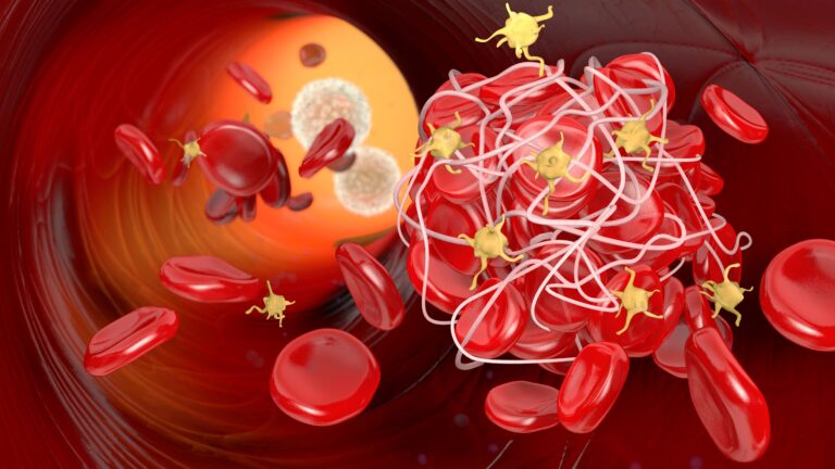
Key Takeaway: The tragic historical legacy and modern pathophysiology of Trousseau Syndrome (Cancer-Associated Thrombosis) underscore a vital rule in internal medicine: an unprovoked, recurrent, or migratory clot (migratory thrombophlebitis) in a patient’s limbs is often not a localized vascular failure, but rather the “smoke from a hidden fire” signaling an occult internal adenocarcinoma. Driven by aggressive molecular mechanisms—such as the tumor-derived release of Tissue Factor-rich microparticles, mucin-induced platelet activation via selectins, and oncogenic MET pathway signaling that paralyzes the body’s natural clot-dissolving systems—these malignancies rapidly induce a systemic hypercoagulable state. Because this cancer-associated thrombosis is characteristically resistant to standard Warfarin therapy, modern diagnostic and treatment guidelines necessitate aggressive systemic screening (via tumor markers, CT, or FDG-PET/CT) alongside prompt anticoagulation utilizing low-molecular-weight heparins or direct oral anticoagulants (DOACs) like Apixaban to extinguish the underlying metabolic fire at its roots.
Introduction: The Bloody Whisper of Distant Organs
Clinical medicine achieves its true power when it approaches the human body not as a chest of isolated drawers, but as an integrated ecosystem where every cell communicates with one another. One of the most insidious scenarios an internist encounters at the clinic desk is when a pathology within an organ system manifests itself for the very first time in an entirely unrelated and distant vascular bed. The most striking, loudest, and historically revered example of this is Trousseau Syndrome, known in modern literature as Cancer-Associated Thrombosis (CAT).
When the patient walks through the clinic door, their complaint is neither weight loss nor unremitting pain; it is simply a painful clot suddenly appearing in the leg or arm, healing, and then migrating to another vein (migratory thrombophlebitis). What appears at first glance to be a simple, localized vascular inflammation can, in reality, be the initial cry of an occult (hidden) malignancy growing silently in the abdomen, lungs, or deep tissues, manipulating the coagulation system like a puppet. In this comprehensive analysis, we will delve deep into the tragic story of a 19th-century physician who discovered this condition within his own body, the molecular mechanisms by which cancer cells sabotage blood fluidity, and the diagnostic and therapeutic strategies used in modern medicine today.
The Most Dramatic Diagnosis in Medical History: The Prophecy of Dr. Armand Trousseau
In the year 1865, Dr. Armand Trousseau, one of the most brilliant clinicians of his era, held the prestigious chair of medicine at the renowned Hôtel-Dieu Hospital in Paris. Trousseau was a physician who placed immense value on bedside examination, fiercely advocating that medicine was not merely a laboratory science, but a profound art of observation. In his clinical lectures published that year, he formulated a sinister correlation that had previously escaped the medical world’s attention: unexplained, spontaneous superficial clotting patterns that migrated from one vein to another (migratory thrombophlebitis) could actually serve as an early and specific harbinger of an occult internal malignancy, particularly adenocarcinomas of the stomach and pancreas. With this groundbreaking observation, Trousseau became one of the founding fathers of the concept of paraneoplastic syndromes.
Yet, destiny had orchestrated an incredibly ironic and tragic script for this brilliant medical genius. Just two years after this landmark publication, in early 1867, Dr. Trousseau noticed a painful, indurated, linear swelling in his own left forearm. While this localized inflammation subsided within a few days, similar vascular cords soon emerged in his other limbs. Trousseau recognized the stealthy phantom he had diagnosed in hundreds of his own patients: migratory thrombophlebitis. Without the need for a single laboratory test or imaging modality, the grandmaster felt the cold breath of his own theory on his neck. He urgently summoned his closest and most devoted student, Dr. Michel Peter, to his chambers. Pointing to the thrombosed vein on his arm, he uttered those historic words that would be etched into medical history with profound sorrow:
“My dear friend Peter, I am lost. A phlebitis that suddenly declared itself last night leaves me with absolutely no doubt regarding the true nature of my illness. My coagulation theory is operating flawlessly this time—upon myself. A hidden tumor is growing within. I wrote my own death warrant with my own hands during my clinical lectures, and now, I am reading it upon my own flesh.”
Dr. Trousseau’s clinical foresight proved devastatingly accurate. Just a few months after diagnosing his eponymous syndrome within his own body, he passed away in June 1867 due to advanced gastric cancer (gastric carcinoma), precisely as his clinical algorithms had predicted. Following his passing, his students Dieulafoy and Peter documented their mentor’s stoic and dignified final days in the medical archives. This clinical event remains recorded in history as the most dramatic and literary self-diagnosis in the annals of medicine.
In summary, Trousseau syndrome is a state of cancer-induced hypercoagulability that manifests as recurrent venous and arterial thromboses, including stroke and pulmonary embolism. It is predominantly associated with advanced, often mucin-producing cancers, revealing itself through migrating or unexplained clots and elevated D-Dimer levels. Early detection, aggressive malignancy screening, and prompt, typically heparin-based anticoagulation management are of central importance. Ultimately, this syndrome stands as a direct systemic reflection of the underlying malignancy’s aggressive biological nature.
The Molecular Matrix: How Cancer Cells Clot the Blood
The molecular mystery behind the syndrome that Dr. Trousseau discerned in the 19th century using only his powers of observation was fully illuminated by 20th and 21st-century hematology and onkoloji research. Cancer is not just an uncontrolled mass of dividing cells; it is also a microscopic factory that drives the systemic circulation into a procoagulant storm. The disruption of the coagulation balance within the extracellular matrix and the vascular bed progresses primarily through three main molecular mechanisms:
Tissue Factor (TF) and Microparticle Bombardment
In a healthy circulatory system, Tissue Factor (TF) resides strictly within the outer layers of the vessel wall (adventitia) to prevent blood from leaking outside the vascular space. However, adenocarcinoma cells—particularly those originating in the pancreas, stomach, lungs, and colorectal regions—reveal abnormally high amounts of functional Tissue Factor on their surfaces. Even more dangerously, these tumor cells shed tumor-derived microparticles (microvesicles) into the bloodstream. These microscopic sacs, coated with active Tissue Factor, travel through the entire circulatory system, continuously triggering the extrinsic pathway mechanism of the coagulation cascade by binding with Factor VIIa. This continuous activation generates a massive, systemic wave of thrombin production.
Mucinous Secretion-Induced “Stickiness” and Selectin Interaction
Adenocarcinomas originating from glandular tissues secrete heavily hydrated protein chains called mucins into the bloodstream. Mucin molecules do not disrupt blood fluidity through direct mechanical obstruction; instead, they execute a biochemical sabotage. Specific oligosaccharide structures on the mucin chains bind to L-selectin on circulating leukocytes (white blood cells) and P-selectin on platelets (clotting cells). This binding aggressively activates the platelets. These activated platelets and leukocytes adhere to one another, forming small cellular clusters (micro-aggregates) within the vessels. These microscopic cellular clusters provide the clearest explanation for why D-Dimer levels continuously rise in cancer patients, even in the absence of a macro-thrombosis.
Cancer Signaling Pathways: MET and Oncogene Activation
In tumor biology, the activation of the MET oncogene simultaneously enhances the cancer’s invasive capability (invasion and metastasis) and directly stimulates the coagulation cascade. Overexpression of the MET signaling pathway upregulates the intracellular production of both Tissue Factor and Plasminojen Activator Inhibitor-1 (PAI-1). The surge in PAI-1 effectively blocks fibrinolysis—the body’s natural breakdown system for dissolving clots. Consequently, the cancer cell heavily accelerates clot production on one hand, while entirely paralyzing the mechanisms the body relies on to dissolve those clots on the other.
“The Smoke in the Chimney and the Hidden Fire Inside the House”
When speaking with anxious patients who present to my outpatient clinic with unexplained, recurrent clotting or highly elevated D-Dimer reports, I always use the following clinical metaphor to help them visualize this insidious condition. Furthermore, this analogy serves as a diagnostic compass for my younger colleagues reading my clinical notes:
“Imagine you are walking past your house. From the outside, everything looks peaceful and quiet; the windows are shut. However, when you lift your head, you notice thick, black smoke pouring out of the chimney. There are no visible flames inside the house, and the fireplace has not been lit. In this situation, can you simply look at the chimney and say, ‘Oh, look how beautifully the smoke rises’? Or can you solve the problem simply by capping the chimney—meaning, giving a quick blood thinner and brushing the clot aside? Absolutely not. If smoke is coming out of that chimney, it tells you with absolute certainty that there is a fire smoldering deep inside the basement, the kitchen, or a hidden room.
The migratory clots (Trousseau Syndrome) that appear out of nowhere in a patient’s leg or arm—without any history of trauma, long-distance flights, or recent surgery—are exactly like the smoke rising from that chimney. The clot itself is not a localized vascular disease; it is merely the smoke generated by a hidden tumor growing silently deep within, setting the coagulation system ablaze. Our duty as clinicians is not merely to extinguish the smoke at the chimney by dissolving the immediate clot; we must trace that smoke to its source and uncover the hidden fire—the occult malignancy. We must manage and treat the underlying insidious disease rather than just chasing the numbers or the localized thrombosis.”
Clinical Manifestations and Diagnostic Approach: Hunting the Hidden Enemy
The clinical spectrum of Trousseau Syndrome is remarkably broad. While its most classic presentation is superficial migratory thrombophlebitis, today this syndrome frequently encompasses deep vein thrombosis (DVT), pulmonary embolism (PE), and even non-bacterial thrombotic endocarditis (NBTE)—characterized by microscopic clot vegetations forming on the heart valves.
- Anticoagulation Resistance: A hallmark clinical feature of these patients is the failure to halt the thrombotic process despite receiving standard doses of conventional blood thinners (such as Warfarin or standard DOACs), with new clots continuously breaking out in different vascular beds. If a patient experiences recurrent thrombosis while undergoing therapeutic anticoagulation, Trousseau Syndrome must be the primary differential diagnosis considered.
- Diagnostic Screening Algorithm: In a patient presenting with an unprovoked, idiopathic thromboembolism where Trousseau syndrome is suspected, malignancy screening must be aggressive. Beyond a complete blood count, routine biochemistry, and serum tumor markers (CA 19-9, CEA, CA-125), a Computed Tomography (CT) scan of the Chest, Abdomen, and Pelvis serves as the first line of defense. If these initial scans fail to identify a primary source but clinical suspicion and D-Dimer elevations persist, a gastrointestinal endoscopy and, if necessary, a whole-body FDG-PET/CT scan should be performed to locate that ‘hidden fire’ Dr. Trousseau warned us about.
Shifting Therapeutic Paradigms: From LMWH to Smart Molecules
The management of cancer-associated thrombosis differs fundamentally from standard clot treatments, a reality that challenged the medical community for decades.
- The Failure of Warfarin (Coumadin): Vitamin K antagonists (Warfarin), which served as the backbone of thrombotic care for generations, are notoriously ineffective in Trousseau Syndrome. Because tumor-derived mucin and tissue factor activate the coagulation cascade directly and independently of the hepatic synthesis of clotting factors, Warfarin cannot reliably suppress recurrent thrombotic events in these patients.
- Low-Molecular-Weight Heparins (LMWH): Landmark clinical trials such as CLOT and CATCH proved that subcutaneous low-molecular-weight heparins, such as Enoxaparin or Dalteparin administered once or twice daily, reduced the rate of recurrent thrombosis by 50% compared to Warfarin, establishing them as the gold standard for years. Heparin does more than just mechanically inhibit thrombin; its molecular structure directly interferes with the binding of tumor cells to selectin receptors, systematically disrupting the core pathophysiology of Trousseau Syndrome.
- The DOAC Revolution (Direct Oral Antikoagülanlar): Recently published clinical data from trials like SELECT-D (Rivaroxaban), Hokusai-VTE Cancer (Edoxaban), and notably CARAVAGGIO (Apixaban) have demonstrated that next-generation direct oral anticoagulants (Factor Xa inhibitors) are just as safe and effective as subcutaneous injections. Today, for cancer patients who do not present with a high risk of gastrointestinal hemorrhage, oral DOAC molecules have rightfully become the frontline therapeutic choice, significantly enhancing patient quality of life.
Conclusion: Looking to the Future in the Light of a Clinical Legacy
Dr. Armand Trousseau presented his own body as a living laboratory to medical science, unveiling the profound and stealthy vascular-oncological links within the human organism. Today, his legacy stands as one of our most potent diagnostic guides across modern hematology laboratories and oncology wards. It must never be forgotten that a clot suddenly appearing within the vascular bed is occasionally far more than a localized mechanical failure; it can be a vital clinical window provided to us to capture a life-threatening enemy early. The true art of medicine lies in the ability to hear the whisper behind that clot.
Our Slogan: “A clot in the vein is sometimes not a local failure, but the smoke from a hidden fire rising through the chimney; monitor the smoke calmly, but work tirelessly to extinguish the fire at its roots.”
Key Clinical Studies & Guidelines Reviewed
- Metharom P, Falasca M, Berndt MC. The History of Armand Trousseau and Cancer-Associated Thrombosis. Cancers (Basel). 2019;11(2):158.
- Wells PS, Anderson DR, Rodger M, et al. Excluding pulmonary embolism at the bedside without diagnostic imaging: management of patients with suspected pulmonary embolism based on clinical probability and D-dimer measurement. Annals of Internal Medicine. 2001;135(2):98-107.
- Kline JA, Courtney DM, Kabrhel C, et al. Clinical criteria to prevent unnecessary diagnostic testing in emergency department patients with suspected pulmonary embolism. Journal of Thrombosis and Haemostasis. 2004;2(8):1247-1255.
- Varki A. Trousseau’s syndrome: multiple definitions and multiple mechanisms. Blood. 2007;110(6):1723-1729.
- Palacios-Acedo A, Langiu M, Crescence L, Mège D, Dubois C, & Panicot-Dubois L. Platelet and Cancer-Cell Interactions Modulate Cancer-Associated Thrombosis Risk in Different Cancer Types. Cancers (Basel). 2022;14(3):730.
- Agnelli G, Becattini C, Meyer G, et al. (Caravaggio Investigators): Apixaban for the Treatment of Venous Thromboembolism Associated with Cancer. The New England Journal of Medicine. 2020;382(17):1599-1607.
Medically reviewed by
Dr. Şekip Altunkan
Dr. Şekip Altunkan is an internal medicine specialist with extensive clinical experience. He trained at Hacettepe University Faculty of Medicine and later served as an Associate Professor in Internal Medicine. He founded and led the Metropol Internal Medicine and Hypertension Clinic in Ankara, pioneering non-invasive Electron Beam Tomography (EBT) cardiac imaging, arterial-stiffness measurement, and nationwide Holter monitoring. He currently practices at his private clinic in Ankara, focusing on hypertension, vascular health, cholesterol, diabetes and heart disease. He has published widely in national and international journals, serves as a peer reviewer for several international journals, and is the author of the book "Questions and Answers on Hypertension."