The Clinical Labyrinth of Coronary Artery Calcium Accumulation in Asymptomatic Patients
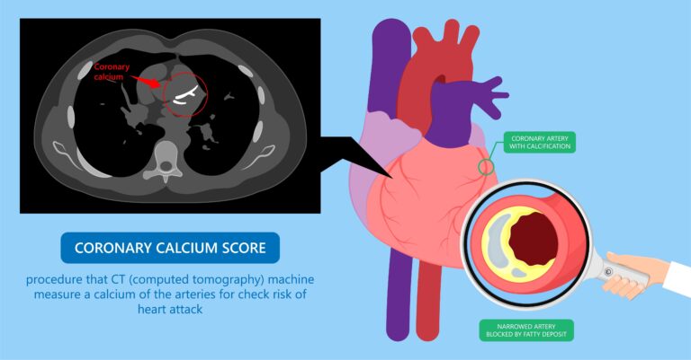
Key Takeaway: Routine Coronary Computed Tomography Angiography (CCTA) performed on completely asymptomatic patients creates a clinical Pandora’s box, often yielding incidental findings that cause profound patient anxiety without providing a survival advantage. When an initial non-contrast scan reveals an advanced Agatston score exceeding the critical threshold of 400—such as a score of 825—international guidelines mandate halting the contrast phase due to the physical limitations of tomographic detectors. At these elevated densities, dense limestone-like calcifications trigger a severe “blooming artifact,” where X-ray attenuation scatters and creates an exaggerated, glowing halo on the screen that misinterprets a non-obstructive 30 percent plaque as an 80 percent critical stenosis. Pathophysiologically, coronary calcium represents a protective, fossilized tombstone of a historical inflammatory war where smooth muscle cells differentiated into osteoblast-like phenotypes to stabilize the arterial matrix against acute rupture. Anchored by the landmark ISCHEMIA trial, optimal clinical management for stable, asymptomatic individuals dictates shelving invasive catheterization in favor of non-invasive functional testing—such as stress echocardiography or myocardial perfusion scintigraphy—paired with aggressive optimal medical therapy, high-intensity statins, and strict blood pressure control to treat the biological reality of the patient rather than the optical illusions of a radiology report.
Introduction
I would like to begin this article with a philosophical premise: “Medicine is a massive detective game predicated on unearthing the flaws of human nature.” Yet sometimes, when no crime has been committed, no glass broken, and no alarm sounded, the detective who enters the crime scene purely out of curiosity can mistake his own footprints for evidence and send an innocent man to prison.
Let me briefly explain why I wanted to write this article: A hypertensive patient presented to our clinic for an evaluation and a second opinion. This 65-year-old male had undergone a CT coronary angiography approximately one month prior, despite having zero cardiac complaints, purely for routine check-up purposes. The outcome was a complete devastation for the patient; his coronary artery calcium score was measured at 825, and during the contrast-enhanced phase, he was told there was severe stenosis in two of his vessels, concluding with an urgent recommendation for invasive angiography. Following this test, the patient lost his sleep, paralyzed by the fear that he could suffer a heart attack at any moment. Now, through the specific lens of this patient, I want to elucidate the true importance of CT coronary angiography and coronary artery calcium scoring in risk stratification.
Driven by the modern era’s desire to “know everything,” performing a Coronary Computed Tomography Angiography (CCTA) as a routine check-up on a completely asymptomatic body is, in reality, a medical Pandora’s box that should remain sealed. And when that box was opened, a mountain of calcium with an Agatston score of 825 and a report claiming “severe stenosis” in two vessels emerged. Here are the two major reasons behind the patient’s sleepless nights.
Now, as both a clinician and a storyteller, I will sit at the table, and we will peel back the pathophysiological realities behind this radiological illusion layer by layer. Are we facing a genuine heart attack risk—metaphorically speaking, a death warrant—or simply a technological optical illusion played by light?
The Cost of Curiosity: Tomographic Intrusion on an Asymptomatic Body
The answer to our first question hits one of the most unyielding walls of clinical cardiology: No, it is fundamentally incorrect to perform an immediate contrast-enhanced Coronary CT Angiography (CCTA) on a patient with no cardiac complaints. This approach is explicitly classified as Class III (harm exceeding benefit) for asymptomatic individuals in the guidelines of both the American College of Cardiology/American Heart Association (ACC/AHA) and the European Society of Cardiology (ESC). Why? Because the silence of the body is evidence of a delicate homeostatic balance maintained at the cellular level. CCTA is an exclusion weapon, exquisitely designed to rapidly rule out coronary disease in patients presenting with suspicious chest pain to the emergency department or those trapped in the gray zone of a treadmill test. However, when deployed in an asymptomatic individual, it unleashes a storm of “incidentalomas”—accidental findings that lack clinical or hemodynamic significance and do not disrupt myocardial perfusion in the slightest. It detects age-related, hypertensive, or genetically driven plaques that have not compromised the coronary architecture even to a minor degree. Consequently, the patient enters the white room believing they are healthy and exits with a report alleging severe blockages, their palms sweating with profound anxiety. This pathway toward the invasive catheterization table leaves the patient exposed to the complications of an entirely unwarranted percutaneous coronary intervention (PCI). Scanning the coronary arteries with tomography during an asymptomatic period is akin to tampering with the mechanism of a perfectly functioning clock out of sheer curiosity, risking throwing its entire alignment out of order. I must, however, exclude the non-contrast calcium score from this restriction. I will elucidate the reasons for this further down in the article.
When Light Deceives: The Blooming Artifact
The phrase “severe stenosis in two vessels” scrawled across a radiology report is not proof of an absolute occlusion within the patient’s heart, but rather evidence that the CT scanner has reached its physical limits. In a patient with a calcium score of 825, the coronary arteries have practically transformed into tubes lined with limestone. At this juncture, when tomographic detectors encounter calcium possessing a high Hounsfield Unit (HU), they lose the ability to accurately interpret X-ray attenuation. To illustrate this phenomenon through a literary lens: Imagine driving down a dark, foggy road at night. An oncoming vehicle approaches with blinding high beams (calcium). The intense light scatters across your visual field and vehicle lens (detector), creating a dazzling, exaggerated halo vastly larger than the car itself. Looking at that glare, you might assume a massive truck is bearing down on you, when in reality it is just a compact automobile. In computed tomography, this is known as the Blooming Artifact. Calcified plaques literally “bloom” on the screen, bleeding over the margins of the dark fluid lumen (the internal cavity). A dense lime layer that actually occupies a mere 30% of the vessel space and leaves blood flow completely unhindered—growing inward into the vessel wall via positive remodeling—projects a pitch-black shadow on screen that mimics an 80% critical obstruction. Consequently, when a scan reads “severe stenosis” in the presence of a score of 825, it represents a massive false-positive. The CT scanner has essentially been blinded by the sheer brilliance of the calcium.
Calcium as a Silent Armor: Was CAC Alone Sufficient?
To quantify the atherosclerotic burden (arterial stiffness) and make an informed decision regarding statin therapy in an asymptomatic individual, a non-contrast Coronary Artery Calcium (CAC – Agatston) scan is more than sufficient. In fact, this has emerged as the gold standard of modern preventive cardiology. Calcium is not the active, malevolent force choking off blood vessels that public perception often assumes it to be. At the pathophysiological level, calcification is the body’s method of laying down concrete to extinguish a cellular fire fueled by cholesterol (LDL) and macrophage-driven inflammation within the arterial wall. Calcium is the tombstone of a historical atherosclerotic war that has been successfully fought and healed. A heavily calcified plaque is profoundly more stable than a soft, lipid-rich plaque, and is highly resistant to acute rupture and subsequent myocardial infarction (MI). Had this patient only received a CAC score—which at 825 indicates a high-risk profile—the clinician’s duty would have been straightforward: pull the prescription pad forward and initiate therapy, without ever injecting iodinated contrast media (dye) or exposing the patient to unnecessary radiation. A score of 825 simply states, “There is an active atherosclerotic process within this patient’s vascular walls.” You initiate a statin, tightly control the hypertension, and send the patient back to their life.
The Threshold of Optical Blindness: Halting the Contrast Phase
According to a universal coronary CT angiography protocol, a patient is first placed in the scanner for a brief, non-contrast scout scan (CAC phase). If the monitor displays an Agatston score exceeding 400 (or >1000 in certain elite centers and next-generation dual-source scanners), international radiology and cardiology guidelines issue a silent mandate to the radiologist: Halt the procedure. This is because once the score breaches the 400 threshold, the sheer density of the calcium renders it virtually impossible to reliably evaluate the transit of contrast material (blood) through the lumen. Injecting an iodinated contrast medium to proceed with a CCTA under these conditions is akin to attempting to photograph a snowman standing against a white wall; the contrast borders dissolve, and margins become entirely obscured. This is why the boundary is strictly set at 400 Agatston units. Beyond this point, the diagnostic specificity of the contrast phase plummets dramatically. The facility evaluating our patient should have capped the needle the moment they witnessed a baseline score of 825.
The Roadmap Forward: Non-Invasive Wisdom at the Crossroads
We are now faced with an asymptomatic patient who has hypertension, a calcium score of 825, and a terrifying report advising him to proceed to invasive angiography. Should our next step be advancing wires through the femoral or radial artery (invasive coronary angiography) to deploy a stent? Absolutely not. At this crossroad, our clinical vision must anchor itself in the paradigm-shifting data of the landmark ISCHEMIA (2020) trial. The ISCHEMIA trial definitively demonstrated that even in the presence of anatomically severe coronary artery disease and associated ischemia, if the clinical picture is stable and free of an acute coronary syndrome, an invasive strategy involving stenting offers no survival advantage or protection against myocardial infarction over optimal medical therapy (OMT) alone.
The management algorithm that should be deployed for this patient rests on this non-invasive wisdom:
- Dynamic Ischemia Evaluation: We must prove whether the “80% tomographic stenosis” has any hemodynamic significance—meaning, whether it is actually depriving the myocardium of blood during stress. The patient must be transitioned to functional testing, such as a Stress ECG, Stress Echocardiography, or Myocardial Perfusion Scintigraphy (MPS).
- Symptom-Objective Alignment: If the stress test or perfusion scan demonstrates completely normal blood flow across the territories supplied by those two suspicious vessels, the CCTA report is discarded as a pure manifestation of the blooming artifact. The recommendation for invasive angiography is permanently shelved.
- The Optimal Medical Therapy (OMT) Armor: Regardless of whether ischemia is present, a calcium score of 825 provides irrefutable evidence of endothelial dysfunction involving the delicate inner lining (intima) of the vascular wall. The patient must be placed under an aggressive, high-intensity statin and ezetimibe regimen. Hypertension must be perfectly regulated to a target of <130/80 mmHg. If clinically indicated, a low-dose aspirin is added to the armor.
We as physicians must resist the temptation to treat the tomography report instead of the patient. After all, calcium is not a fatalistic destiny; rather, it should be evaluated as a protective cellular defense mechanism. We are obligated to enlighten and manage this patient not through invasive means, but through correct medications that stabilize cellular metabolism, combined with composed clinical intelligence.
While major current guidelines do not recommend the calcium score as a universal screening tool for the general population due to evolving cost-effectiveness data , it serves as a powerful “risk marker” in our routine practice to practice industrialized medicine, tailoring therapies for intermediate-risk asymptomatic individuals or those with a premature family history of coronary artery disease.
Molecular Footnotes and Pristine Evidence
Pathophysiology of Calcification: Oxidized LDL particles accumulating within the tunica intima are engulfed by macrophages, transforming them into foam cells. Following this inflammatory battle, these cells undergo apoptosis (programmed cell death), forcing macrophages and smooth muscle cells to differentiate into osteoblast-like phenotypes within the vascular matrix, leading to the precipitation of calcium phosphate (hydroxyapatite) crystals. This molecular ossification acts as a protective phenomenon that stabilizes the plaque architecture and shields it from mechanical rupture. The score of 825 visualized on the screen represents the fossilized remnants of an epic cellular war waged over decades.
CAC continues to serve as a robust indicator of coronary atherosclerosis. Nevertheless, in daily clinical practice, an elevated coronary calcium score (Agatston) may not necessarily signify an active disease state poised for acute rupture. If this elevated score is driven by high calcium density stemming from prolonged statin therapy, advanced age, or a rigorous history of physical endurance exercise, it could instead symbolize advanced plaque stability. In the future, evaluating calcium density as a continuous variable rather than utilizing rigid, discrete Agatston categories, or incorporating an “average density” concept into risk calculators, will provide clinicians with highly sophisticated diagnostic guidelines in both primary and secondary prevention. Research into these methodologies is ongoing. Furthermore, investigations regarding artificial intelligence-integrated calcium scoring are actively underway. The findings derived from these studies will equip clinicians with powerful diagnostic weapons.
In conclusion, to resolve our patient’s profound distress, I explained a comprehensive summary of these precise principles. We then performed a Myocardial Perfusion Scintigraphy (MPS) study. Once the scan returned entirely normal, alongside medications to meticulously control his hypertension, I prescribed a statin, ezetimibe, and acetylsalicylic acid (Aspirin), placing him under close clinical surveillance. Furthermore, I explained to the patient that this evaluation was far from a waste of time; by uncovering his calcium score, we successfully stratified his long-term risk and tailored an appropriate treatment regimen. I advised him to view this structural clarity as a significant clinical gain.
Let us define our slogan: “The silence of a symptom must never be sacrificed to the optical noise of an image; for an excellent physician treats the reality of the heart before him, not the shadows on a CT report.”
Key Clinical Studies & Guidelines Reviewed
- Maron, D. J. et al. Initial Invasive or Conservative Strategy for Stable Coronary Disease (ISCHEMIA Trial). New England Journal of Medicine. 2020; 382(15), 1395-1407.
- Gulati, M, et al. 2021 AHA/ACC/ASE/CHEST/SAEM/SCCT/SCMR Guideline for the Evaluation and Diagnosis of Chest Pain. Circulation, 2021144(22), e368-e454.
- Hecht, H. S., et al. 2016 SCCT/STR guidelines for coronary artery calcium scoring of noncontrast noncardiac chest CT scans. Journal of Cardiovascular Computed Tomography.2017; 11(1): 74-84.
- Greenland, P et al. Coronary Calcium Score and Cardiovascular Risk. Journal of the American College of Cardiology. 2018; 72(4), 434-447.
- Razavi A, Agatston A, Shaw L, et al. Evolving Role of Calcium Density in Coronary Artery Calcium Scoring and Atherosclerotic Cardiovascular Disease Risk. JACC: Cardiovascular Imaging. 2022;15(7), 1234-1248.
- Charalambous M. A, Filler R, Soteriades EH, et al. (2026). Coronary Artery Calcium Score as a Predictor of Cardiovascular Disease Events and Mortality Among Asymptomatic Working-Age Adults: A Systematic Review. Cureus. 2026;18(3), e101388.
Medically reviewed by
Dr. Şekip Altunkan
Dr. Şekip Altunkan is an internal medicine specialist with extensive clinical experience. He trained at Hacettepe University Faculty of Medicine and later served as an Associate Professor in Internal Medicine. He founded and led the Metropol Internal Medicine and Hypertension Clinic in Ankara, pioneering non-invasive Electron Beam Tomography (EBT) cardiac imaging, arterial-stiffness measurement, and nationwide Holter monitoring. He currently practices at his private clinic in Ankara, focusing on hypertension, vascular health, cholesterol, diabetes and heart disease. He has published widely in national and international journals, serves as a peer reviewer for several international journals, and is the author of the book "Questions and Answers on Hypertension."