The Silent Strokes Hiding in Plain Sight
Key Takeaway: In more than half of patients with cerebral amyloid angiopathy—a common age-related cerebrovascular disease—advanced MRI reveals evidence of new, clinically silent strokes. These ‘incidental’ lesions are not benign findings; they are associated with elevated blood markers of nerve damage and appear to represent active, ongoing disease.
The Strokes No One Feels
Imagine a stroke occurring inside your brain right now—tiny, painless, and so imperceptible that you and everyone around you fail to notice. There is no facial droop, no slurred speech, no rush to the emergency room. You simply continue with your day as if nothing happened. Yet deep within the tissue of your brain, a small cluster of neurons has just lost its blood supply and quietly died. Now, imagine this happening not just once, but repeatedly over months and years. This is the reality for a surprising number of people living with diseases that attack the brain’s smallest blood vessels—and until recently, these events were often dismissed as background noise on imaging scans.
A new study directly challenges this assumption, providing evidence that these so-called “incidental” bright spots on advanced brain MRIs are far from random. They are, instead, the footprints of an active disease process washing over the brain from the inside out.
What Did the Researchers Do?
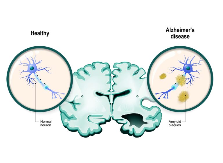
The study examined two separate groups of patients, each suffering from a different type of cerebral small vessel disease. The first group consisted of 43 patients with a diagnosis of probable cerebral amyloid angiopathy (CAA). CAA is a condition in which a sticky protein called amyloid-beta accumulates in the walls of the brain’s small blood vessels, making them fragile and prone to bleeding[2]. CAA is strongly associated with aging and is found in a significant proportion of older individuals, particularly those with Alzheimer’s disease[3]. The second group included 75 patients with CADASIL (cerebral autosomal dominant arteriopathy with subcortical infarcts and leukoencephalopathy), a hereditary condition caused by mutations in the NOTCH3 gene that leads to progressive thickening and degeneration of small artery walls[4].
The researchers employed diffusion-weighted imaging (DWI), a specialized MRI technique that is exceptionally sensitive to the movement of water molecules in tissue. When a stroke occurs, even a microscopic one, cells swell and water movement becomes restricted, creating a distinct bright signal on DWI that standard MRI sequences often miss[5]. The team then compared these imaging findings with blood levels of neurofilament light chain (NfL), a protein released into the bloodstream when nerve fibers are damaged or destroyed[6].
What the Study Found
The results were striking. A full 56% of the 43 CAA patients had incidental DWI-positive lesions—the bright spots indicating new, small, silent strokes[1]. In the CADASIL group, 21% showed the same type of lesions. These were not the strokes that brought the patients to the hospital; they were discovered incidentally during research scans.
Perhaps most illuminating was where these lesions appeared. In the CAA patients, 65% of the lesions were located in the cortex, the outer layer of the brain responsible for high-level thought, language, and perception. In CADASIL patients, 95% of the lesions were found in the subcortical white matter, the deeper wiring that connects different brain regions. This geographical specificity mirrors the known biology of each disease: the amyloid deposits in CAA preferentially affect cortical vessels, while the arteriopathy of CADASIL targets the deeper penetrating arteries.
Crucially, in both patient groups, the presence of these silent lesions was significantly associated with higher serum NfL levels. This is the biological confirmation that transforms an imaging anomaly into a clinical alarm. Elevated NfL means that neurons are being actively damaged. The bright spots are not old scars from the distant past but evidence of a disease in motion.
The Mechanism: Why Small Vessels Cause Major Problems
The brain’s small blood vessels are architectural marvels. Arterioles as thin as a human hair penetrate deep into the brain tissue, delivering oxygen and glucose to neurons that, despite making up only 2% of the body’s weight, consume about 20% of its total energy[7]. These vessels also form the blood-brain barrier, a selective filter that maintains the brain’s delicate chemical environment.
When diseases like CAA or CADASIL compromise these vessels, the consequences cascade. In CAA, the amyloid protein replaces the smooth muscle cells in the vessel walls, turning flexible arterioles into stiff, brittle tubes. Blood flow regulation becomes impaired. Small patches of downstream tissue are starved of oxygen—not dramatically enough to cause symptoms, but sufficiently to kill neurons. In CADASIL, the mutant NOTCH3 protein accumulates in vessel walls, triggering the progressive loss of smooth muscle cells and a narrowing of the vessel lumen[4]. The result is the same: small areas of brain tissue silently infarct, one after another.
What makes this particularly insidious is the cumulative nature of the damage. A single microscopic stroke may be unnoticeable. But dozens or hundreds of them, scattered across critical brain networks over years, can manifest as cognitive decline, gait instability, mood changes, and ultimately, vascular dementia[8]. By the time symptoms become clinically apparent, the accumulated damage may be substantial.
Conclusion: What the Findings Mean for the Future
This study reframes the clinical significance of incidental DWI lesions in patients with known small vessel disease. These findings are not noise to be dismissed in a radiology report but are quantifiable indicators of active brain injury, confirmed by a blood biomarker that independently validates neuronal damage.
For patients with CAA or CADASIL, this has practical implications. First, it suggests that DWI-positive lesions could serve as a way to monitor disease activity over time, much like tracking tumor markers in oncology. Second, it provides a potential endpoint for clinical trials of new therapies targeting small vessel disease. If a treatment reduces the frequency of these silent lesions and lowers NfL levels, researchers would have objective evidence that the intervention is protecting the brain.
For the broader patient population with vascular risk factors—hypertension, diabetes, atrial fibrillation—this research underscores the importance of aggressive vascular risk management. Although this study focused on specific diseases, the principle applies generally: the brain’s small vessels are vulnerable, and damage to them accumulates silently long before it announces itself with a clinical stroke.
Several important caveats exist. The study’s sample sizes were modest—43 CAA patients and 75 CADASIL patients—and the cross-sectional design offers only a snapshot in time. Larger, longitudinal studies are needed to determine if the frequency of these silent lesions predicts long-term cognitive outcomes or responds to treatment. Additionally, the findings apply specifically to patients with established diagnoses of small vessel disease and should not be uncritically generalized to the general population.
Nevertheless, the message is clear and compelling: when a bright spot appears on an advanced MRI of a patient with small vessel disease, it deserves attention, not a footnote. That tiny signal may be the most honest indicator of what is happening inside the brain—a whisper of damage that, if heard early enough, might finally be met with an intervention. However, numerous constraints hinder the widespread use of MRI in clinical practice. Chief among these are the insufficient number of machines and specialists, as well as the high cost. Still, the appearance of such lesions in patients undergoing MRI for any reason is a serious alarm. It necessitates a concerted effort to combat the chronic risk factors that could be causing them.
Scientific Sources
- Ter Telgte A, et al. Incidental DWI-Positive Lesions in 2 Cohorts of CAA and CADASIL: Prevalence, Distribution, and Associations With Clinical Variables. Neurology. 2026;107(2):e218160. PubMed: https://pubmed.ncbi.nlm.nih.gov/42361280/
- Samarasekera N. The association between cerebral amyloid angiopathy and intracerebral haemorrhage: systematic review and meta-analysis. J Neurol Neurosurg Psychiatry. 2012. PMID: 22056966
- Viswanathan A, et al. Cerebral amyloid angiopathy in the elderly. Ann Neurol. 2011. PMID: 22190361
- Chabriat H, et al. CADASIL. Lancet Neurol. 2009. PMID: 19539236
- Schaefer PW, et al. Diffusion-weighted MR imaging of the brain. Radiology. 2000. PMID: 11058626
- Khalil M, et al. Neurofilaments as biomarkers in neurological disorders. Nat Rev Neurol. 2018. PMID: 30171200
- Raichle ME, et al. Brain work and brain imaging. Annu Rev Neurosci. 2006. PMID: 16776593
- Pantoni L. Cerebral small vessel disease: from pathogenesis and clinical characteristics to therapeutic challenges. Lancet Neurol. 2010. PMID: 20610345
Medically reviewed by
Dr. Şekip Altunkan
Dr. Şekip Altunkan is an internal medicine specialist with extensive clinical experience. He trained at Hacettepe University Faculty of Medicine and later served as an Associate Professor in Internal Medicine. He founded and led the Metropol Internal Medicine and Hypertension Clinic in Ankara, pioneering non-invasive Electron Beam Tomography (EBT) cardiac imaging, arterial-stiffness measurement, and nationwide Holter monitoring. He currently practices at his private clinic in Ankara, focusing on hypertension, vascular health, cholesterol, diabetes and heart disease. He has published widely in national and international journals, serves as a peer reviewer for several international journals, and is the author of the book "Questions and Answers on Hypertension."